
PneumaView 3D™
PneumaCare’s PneumaView 3D™ software provides the operator with the ability to divide the image into defined regions for comparison of the patient’s symmetry of breathing. These regional contributions can be analysed by comparing the upper chest and abdomen or the left and right sides of the chest; detailing potential asymmetries that are indicative of respiratory-related issues.
Healthy Subjects
Healthy 19 Year Old Male

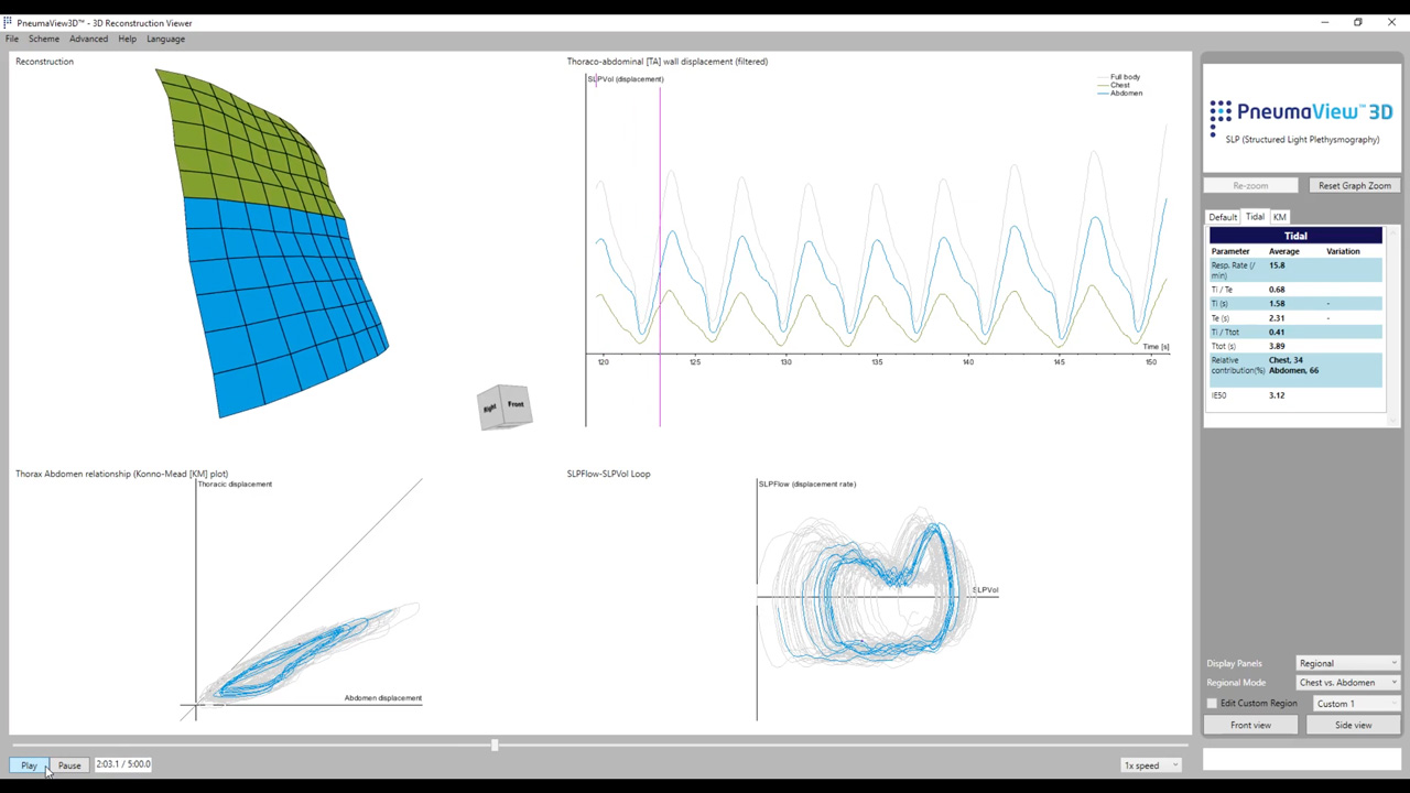
Tidal breathing pattern of a healthy adult measured with Thora-3Di®. The Konno-Mead plot (bottom left figure) appears more or less like a straight line and lies along the 45-degree line and the surrogate flow-volume loop resembles a circle.
Healthy Infant

Thora-3Di® is capable of measuring the tidal breathing pattern in infants and newborn. Infants commonly exhibit a very irregular breathing pattern due to their plasticity. Normal respiratory rates are very high in infants when compared to adults. We have a set of preliminary normative values for children and adults to (2 to 75 years).
COPD Female
Severe High IE50 measurement

Thoraco-abdominal motion of a female patient with severe COPD is captured using Thora-3Di®. IE50 measurement is extremely high, suggesting significant airway obstruction. Thoraco-abdominal asynchrony is apparent in the Konno-Mead plot (bottom left figure) and duty cycle (Ti/Ttot) is lower than the lower limit of normal. The surrogate flow-volume loop appears “squashed” on top suggesting expiratory flow limitation.
Adult with Neuromuscular Disorder
Duchenne Muscular Dystrophy

The scan shows the ability of Thora-3Di® to measure abnormal (deformed) thoraco-abdominal structures. Additionally, respiratory rate is elevated and both inspiratory and expiratory times are abnormally short. IE50 measurement is also below the limit of normal. The Konno-Mead plot (bottom left figure) shows a curved pattern which is rarely seen elsewhere and the surrogate flow-volume loop (bottom right figure) shows higher levels of expiratory flow than inspiratory flow which may indicate inspiratory flow limitation.
Stroke Patient
Double Expiratory Peak
Tidal breathing pattern of a stroke patient measured with Thora-3Di®. The most striking pattern can be seen on the surrogate flow-volume loop on the bottom right. The expiratory portion of the loop peaks twice, which is highly unusual, interestingly, the more dominant expiratory peak occurs towards the end of expiration. Additionally, IE50 measurement is high suggesting airway obstruction and there is a degree of thoraco-abdominal asynchrony shown by the loop generated on the Konno-Mead plot (bottom left figure).
Infant with Acute Viral
Bronchiolitis

An infant with acute viral bronchiolitis measured with Thora-3Di®. Contrary to the tidal breathing patterns expected in infants (i.e. highly irregular), the tidal breathing pattern for this patient appears to be highly regular. Regularity of the tidal breathing pattern can be a sign of disease. There is also a degree of asynchrony between chest and abdominal motion (manifested as a loop in the Konno-Mead plot on the bottom left figure).
Asthma Acute 4 Year Old Male Evident Paradoxical Breathing Pre Bronchodilator

The smaller grid size indicates the ability of Thora-3Di® to measure younger (smaller) subjects, in this case a 4-year male suffering from acute asthma. The paradoxical breathing pattern (chest expanding whilst abdomen simultaneously retracting) is clearly visible on the tidal breathing pattern (top right figure). The Konno-Mead plot (bottom left figure) has formed loops suggesting the same thoraco-abdominal asynchrony (TAA). In fact, when quantified, breath phase of TAA is approximately 120 degrees, which is significantly higher than the upper limit of normal. IE50 measurement is also elevated suggesting a degree of airway obstruction. After administration of a bronchodilator, breath phase for this patient dropped to 56 degrees, whilst still abnormal, this reduction in phase confirms the effect of the intervention.