Our Technology
Standard assessments require a patient to blow vigorously into a small tube while airflow is measured (Spirometry) or to be fitted with a mask which itself can interfere with natural breathing. PneumaCare’s approach addresses this by allowing the lung function assessment to be carried out during natural breathing. PneumaCare offers an alternative.

Spirometry requires a patient to be awake, cooperative, and able to follow instructions; it is therefore difficult to perform with children. Moreover, infants cannot be measured or monitored without direct contact. Data from spirometry provides important clues to help distinguish obstructive pulmonary disorders that typically reduce airflow, such as asthma and emphysema. In addition, spirometry also provides indications regarding restrictive disorders that typically reduce total lung volumes, including pulmonary fibrosis and neuromuscular disease.
Current approaches to lung monitoring have significant shortcomings:
- Can only be used in sub-populations
- Cannot be used in critically ill patients and
- Carry the risk of cross infection.
Due to these shortcomings, because of the considerable physical effort that is required by the patient in blowing into current instruments, up to one third of patients who could benefit from assessment are inaccessible with current techniques. Add to this running cost (from daily calibration, sterilisation, and consumables) and there are considerable limits to the clinical utility of existing market offerings.
Clinicians have voiced a need for an approach that offers faster screening, access to a broader patient population, and dramatically reduces the risk of a hospital-acquired infection. In this current climate the need for a non-aerosol generating and non-contact procedure to aid in treatment pathways.

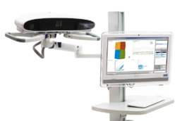
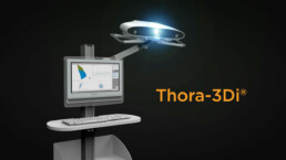
PneumaCare's patented technology is called Structured Light Plethysmography (SLP). This is fully integrated in an easy to use architecture comprising hardware and software.

A grid pattern is projected onto the patient’s chest. Projected light consists of visible white light only, not infrared or UV, with no harm to the patient.
Two cameras film the movement of the grid pattern at high speed (30 frames per second) due to breathing over time.
Software utilizes video to create a 3D view of chest wall movement over time and calculates volume of air moved.
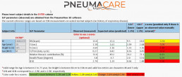
Output is delivered in 3D regional output on the user interface, and complex clinical respiratory parameters are calculated for the physicians use

